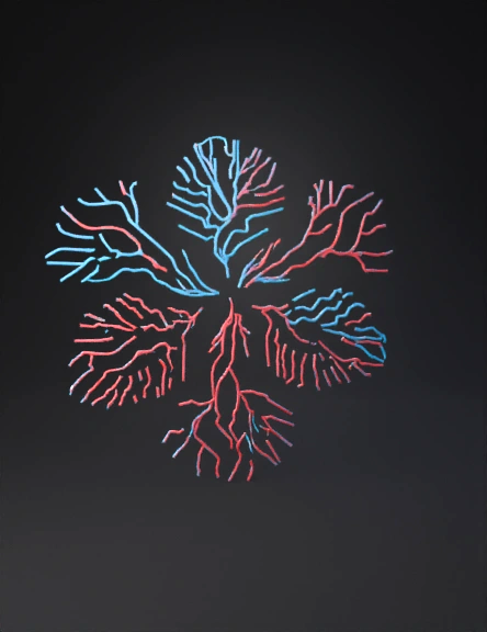
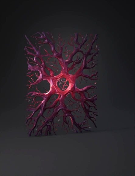
Vascular Network 3D Model
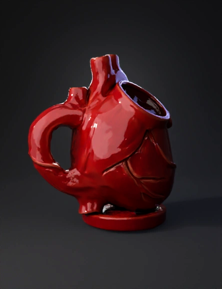
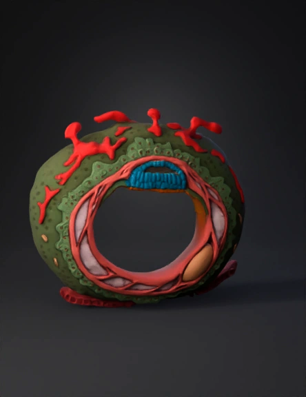
Heart Shaped Vase 3D Model
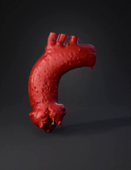
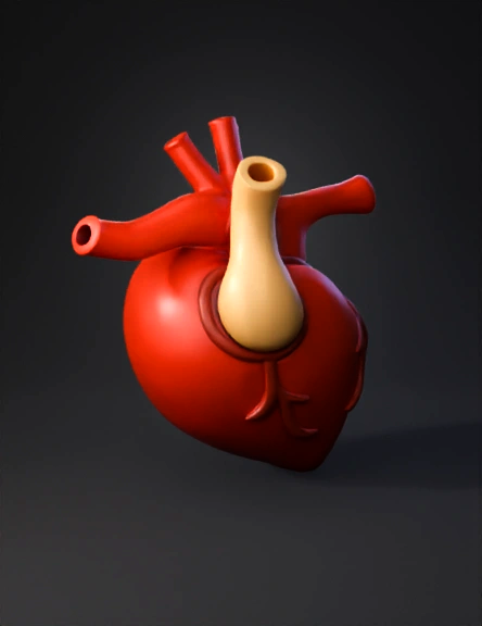
Aorta 3D Model
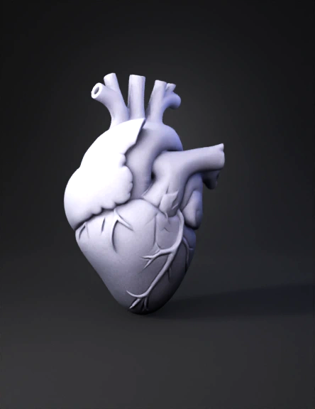
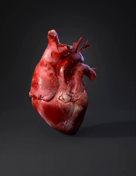
Red Heart 3D Model
Blood Vessel Cross-section 3D Model
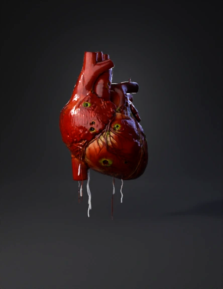
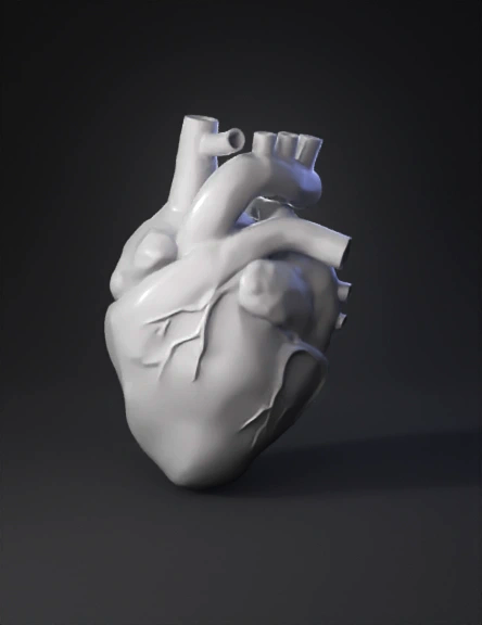
Anatomical Heart 3D Model
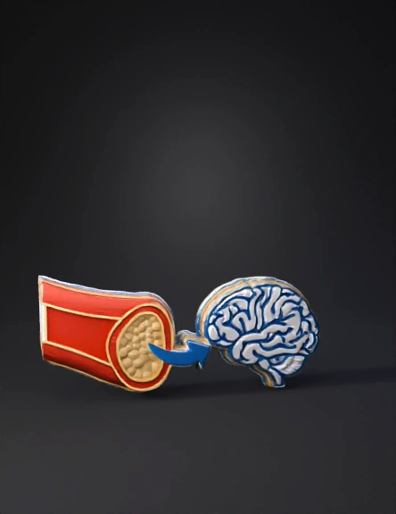
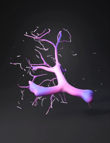
Brain Artery 3D Model
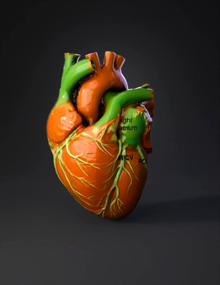
Anatomical Heart 3D Model

Branching Vascular Tree 3D Model
Anatomical Heart 3D Model
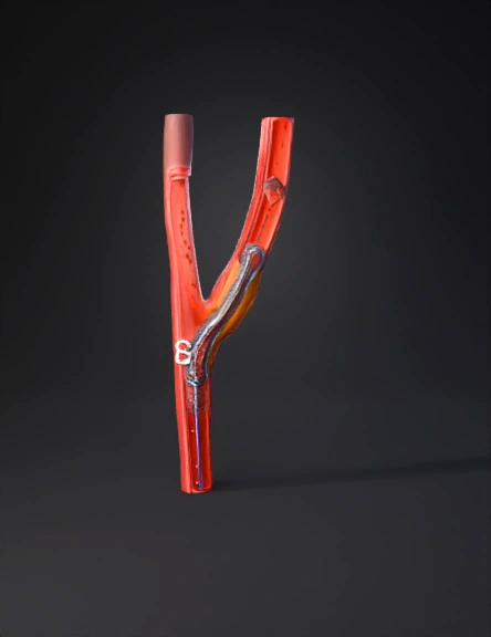
Carotid Artery Stent 3D Model
Vascular Tree 3D Model
Anatomical Heart 3D Model
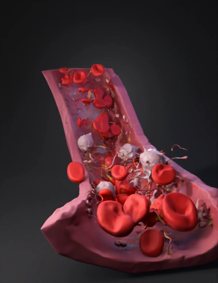
Blood Vessel 3D Model
Modelo 3d De Corazón 3D Model
血管网络三维模型 3D Model
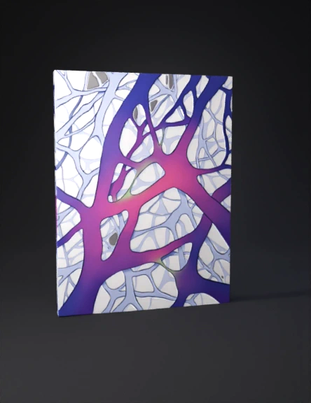
血管 网络 三维 模型 3D Model