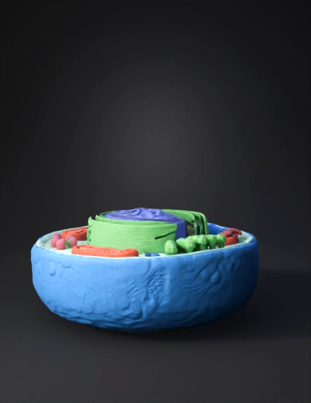
Modelo 3d De Célula Animal 3D Model
Modelo 3d De Célula Procariota 3D Model
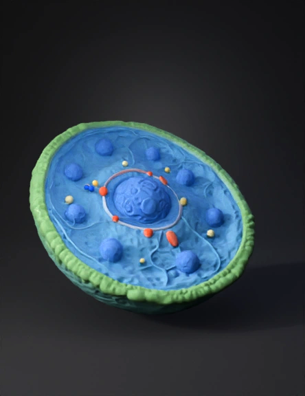
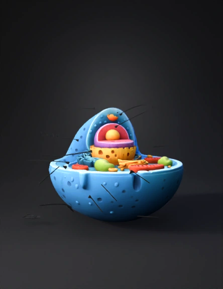
Célula Eucariota 3d 3D Model
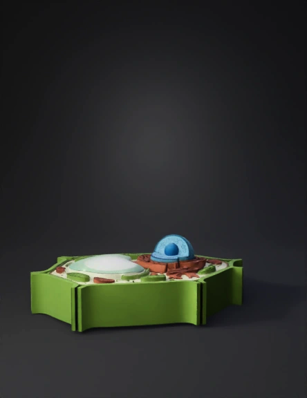
Plant Cell 3D Model
Celula Eucariota 3d 3D Model

Plant Cell 3D Model
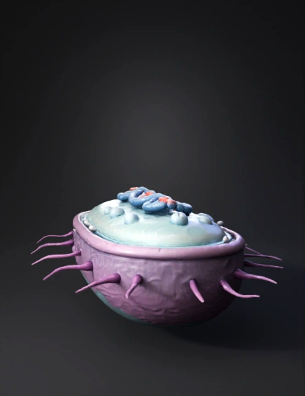
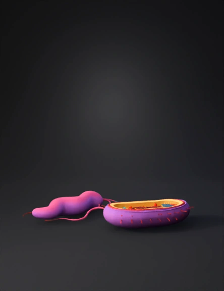
Bacterium 3D Model
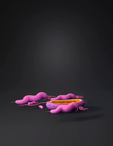
Bacteria Cell 3D Model
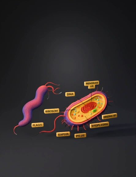
Bacteria Diagram 3D Model
Cell Model 3d 3D Model
Cell Model 3d 3D Model
Cell Model 3d 3D Model
Cell Model 3d 3D Model
Ciliated Cell 3D Model
Biological Cell 3D Model
三维 细胞 模型 3D Model
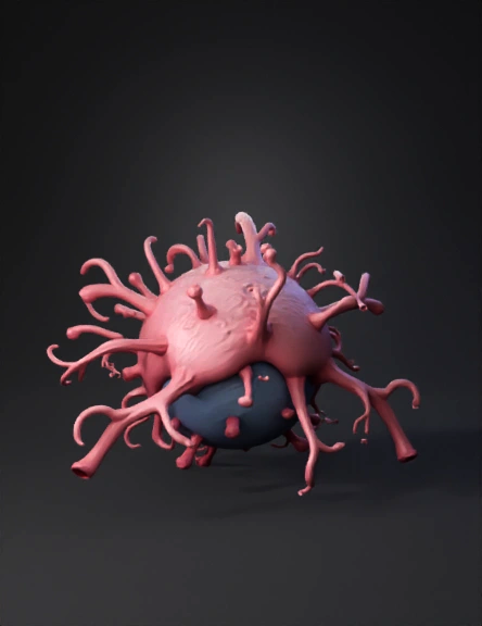
Bacteria 3D Model
Cell Diagram 3D Model