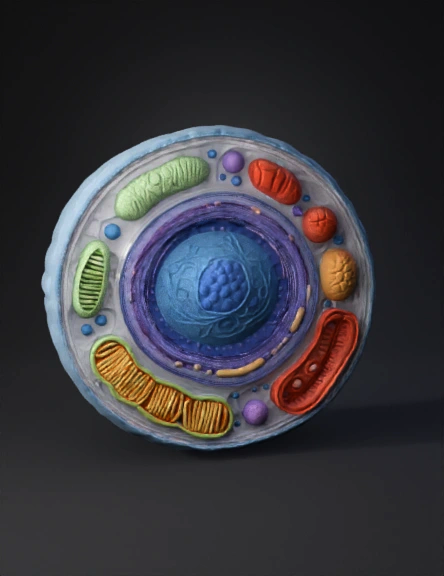
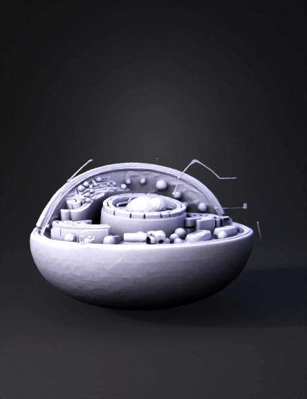
Cell Organelle 3D Model
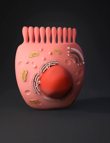
Enterocyte 3D Model
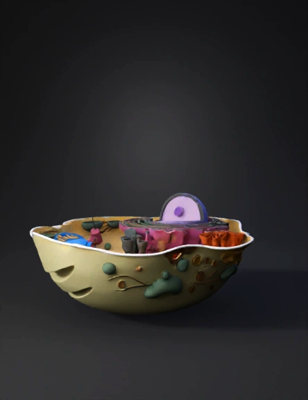
Cell Anatomy 3D Model
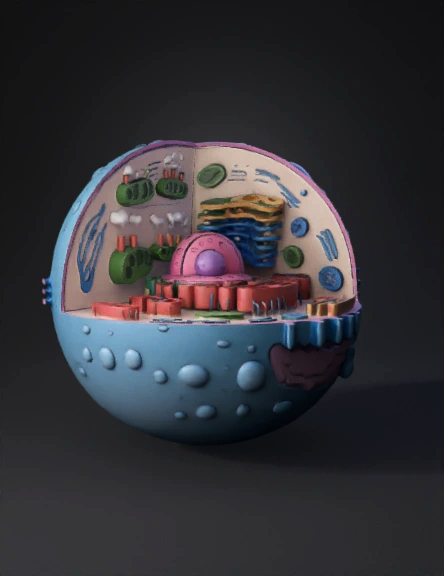
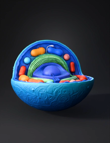
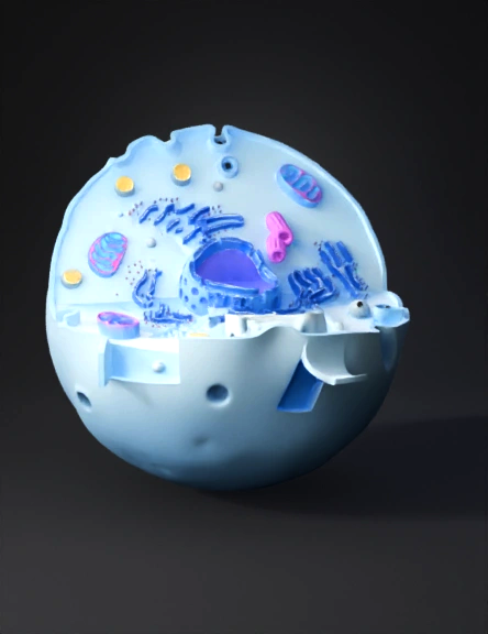
Eukaryotic Cell 3D Model
Cell Structure 3D Model
Cell Anatomy 3D Model
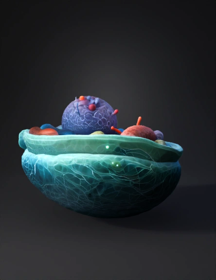
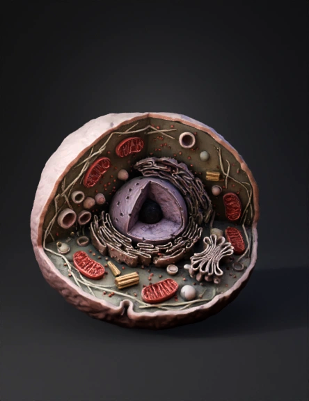
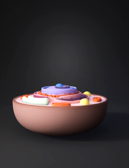
Cell Cross-section 3D Model

Cell Anatomy 3D Model
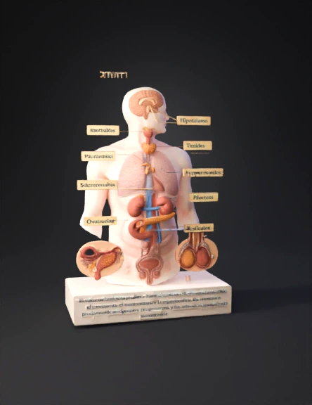
Endocrine System 3D Model
Animal Cell 3D Model
Cell Anatomy Diagram 3D Model
Eukaryotic Cell 3D Model
Animal Cell 3D Model
Cell Diagram 3D Model
Cell Model 3d 3D Model
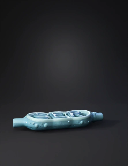
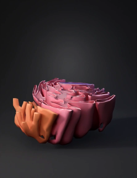
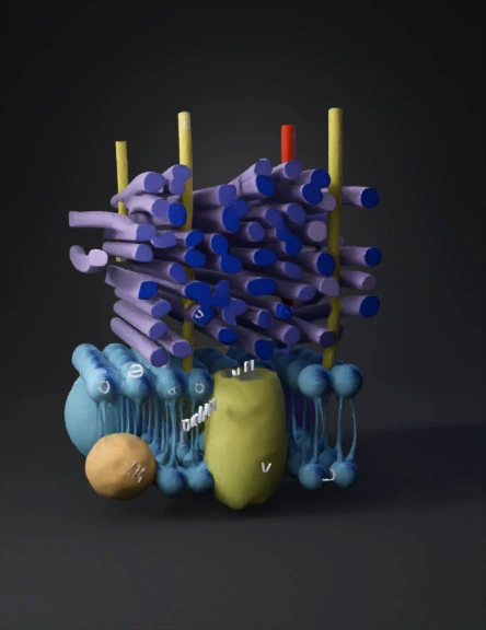
Smooth Endoplasmic Reticulum 3D Model
Cell 3D Model

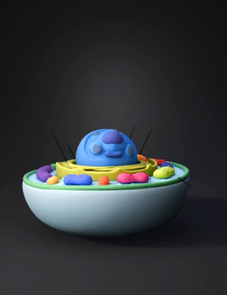
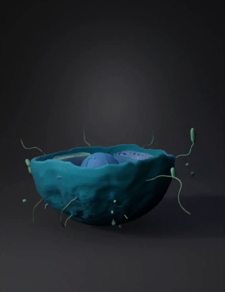
Stylized Cell 3D Model