
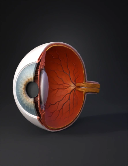
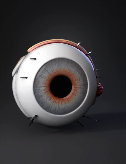
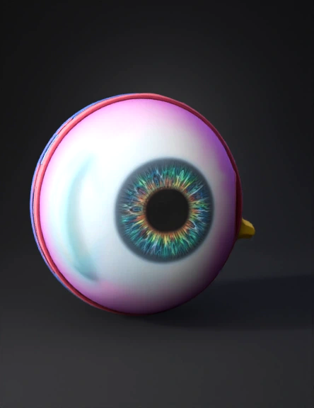
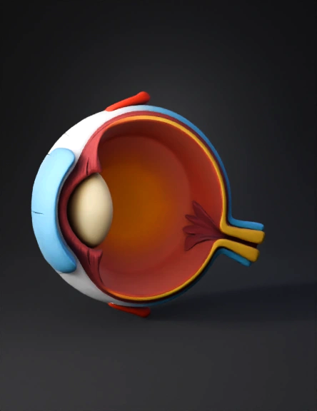
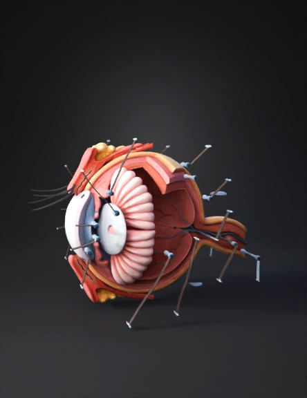
Eye Anatomy 3D Model

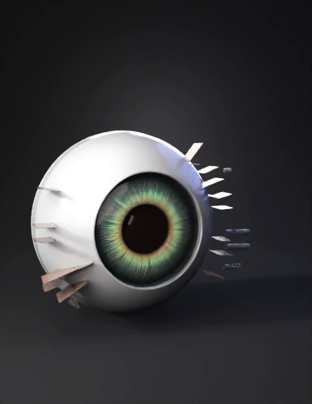
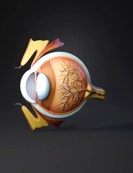
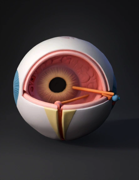
Eye Anatomy 3D Model

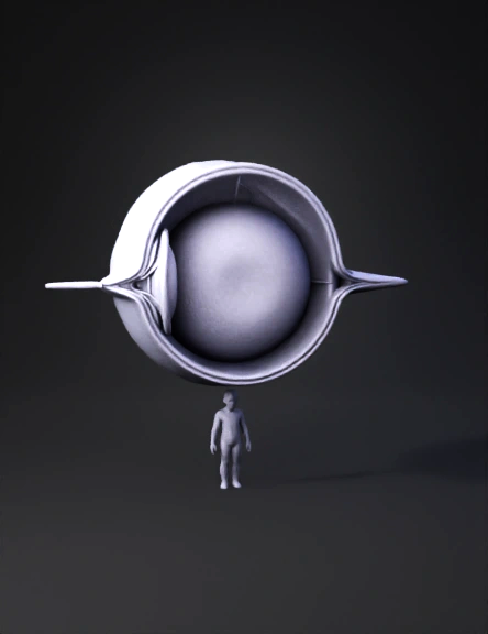
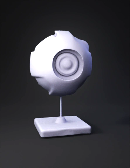
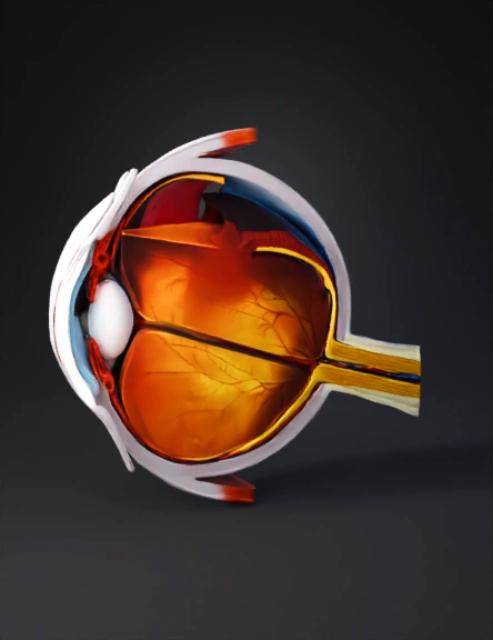
Eye Anatomy 3D Model
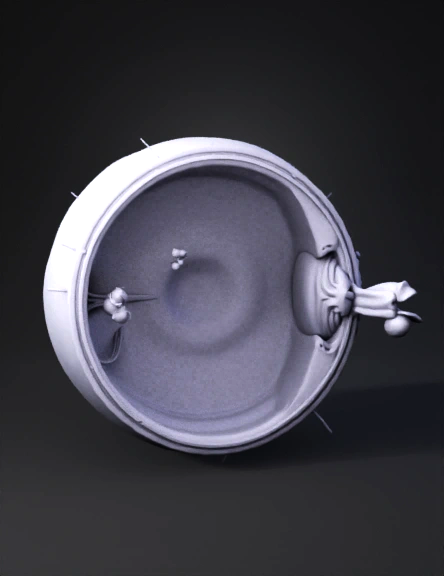
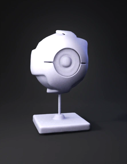
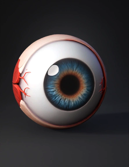
Eye Anatomy 3D Model
Eye Anatomy 3D Model

Eye Anatomy 3D Model
Eye Anatomy 3D Model
Eye Anatomy 3D Model
Eye Anatomy 3D Model
Eye Anatomy 3D Model
Eye Anatomy 3D Model
Eye Anatomy Model 3d 3D Model
Eye Anatomy 3D Model
Eye Anatomy 3D Model
Eye Anatomy 3D Model
Eye Anatomy 3D Model
Eye Anatomy 3D Model
Eye Anatomy 3D Model