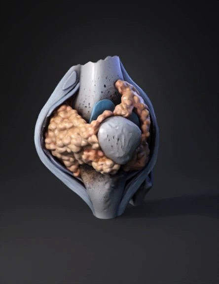
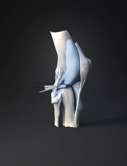
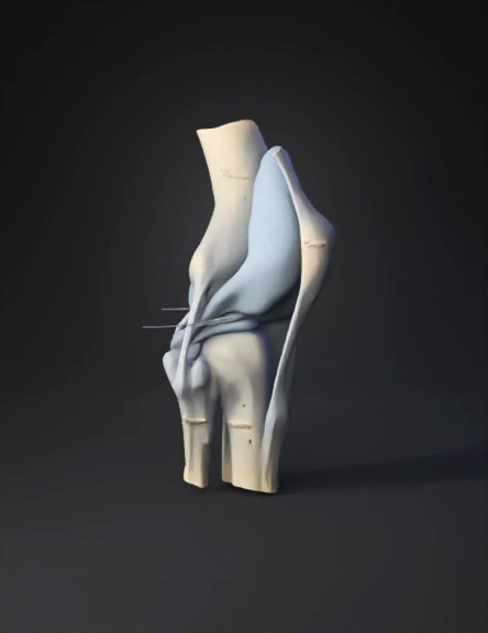
Knee Joint 3D Model
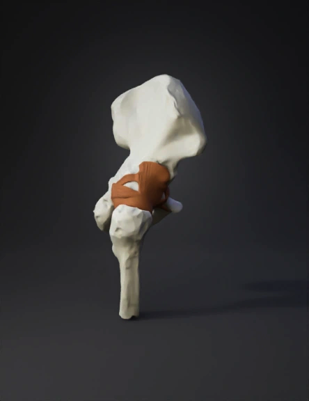
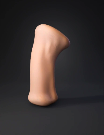
Hip Joint 3D Model
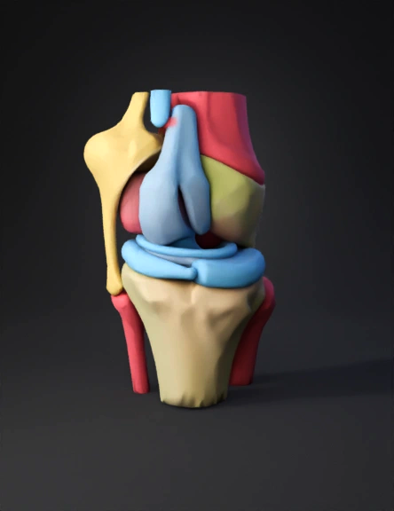
Knee Joint Anatomy 3D Model
Knee Joint Anatomy 3D Model
Knee Joint 3D Model
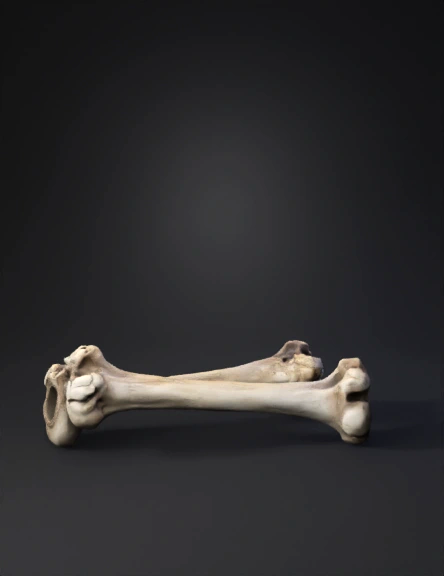
Fémur Modelo 3d 3D Model
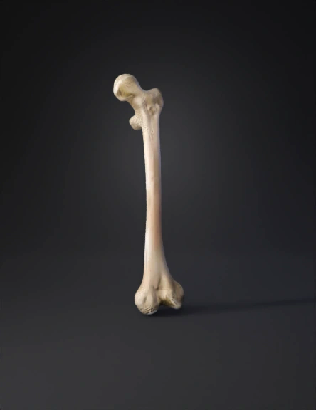
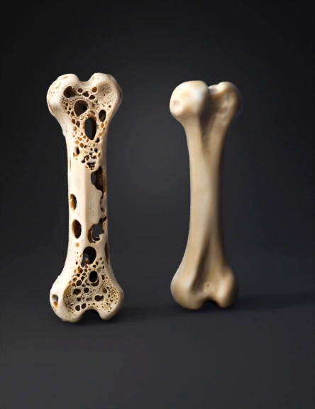
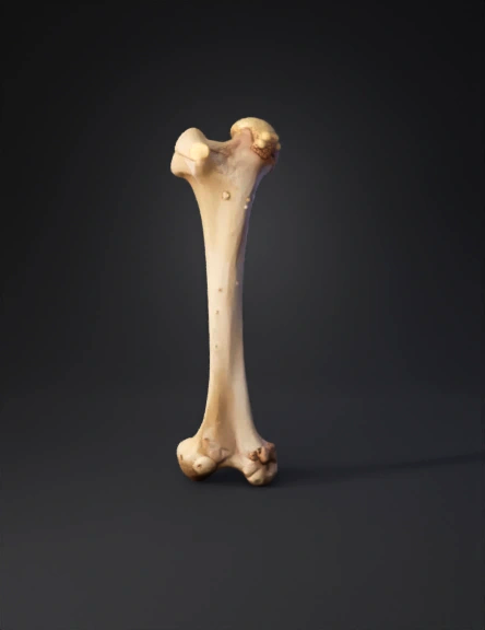
Femur Bone 3D Model

Femur Bone 3D Model
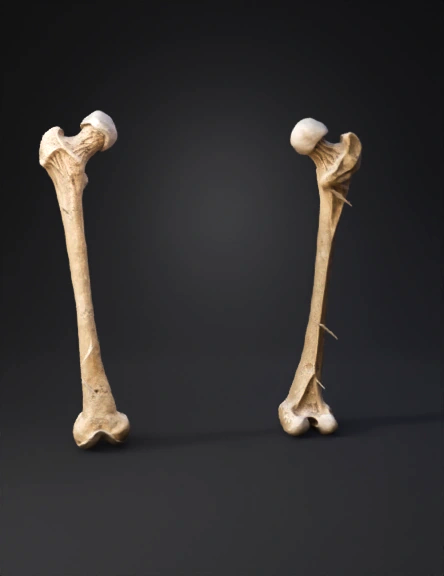
Femur Bone 3D Model
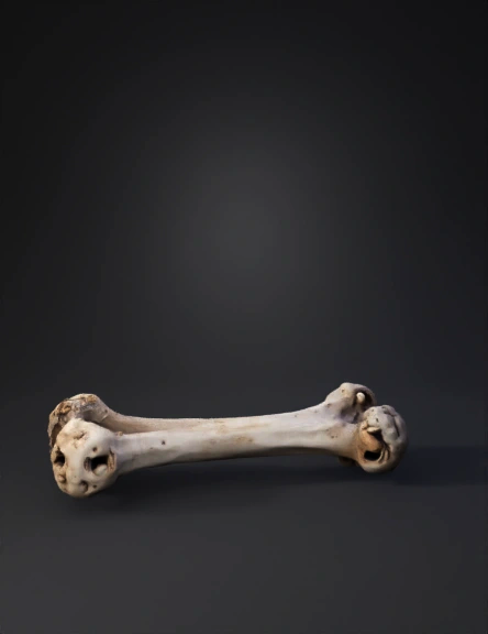
Femur Bone 3D Model
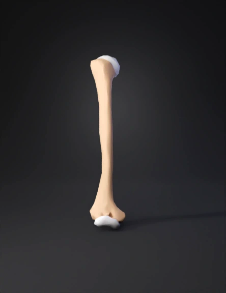
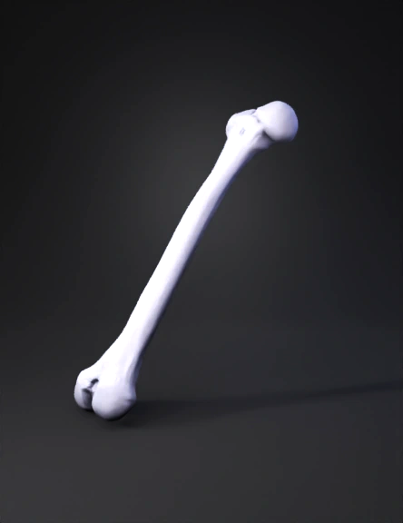
Human Femur Bone 3D Model
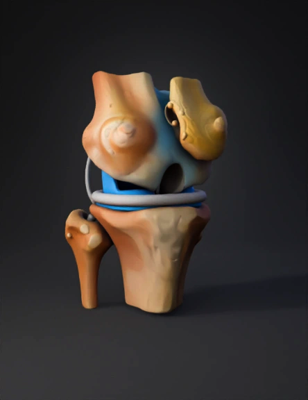
膝关节 3d 模型 3D Model
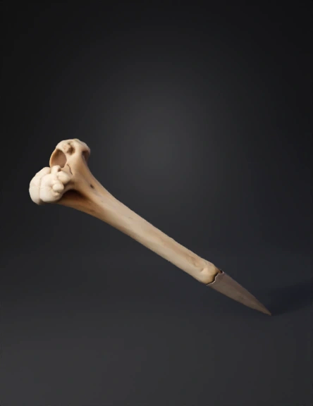
Bone Knife 3D Model
Bone 3D Model
Femur Bone 3D Model
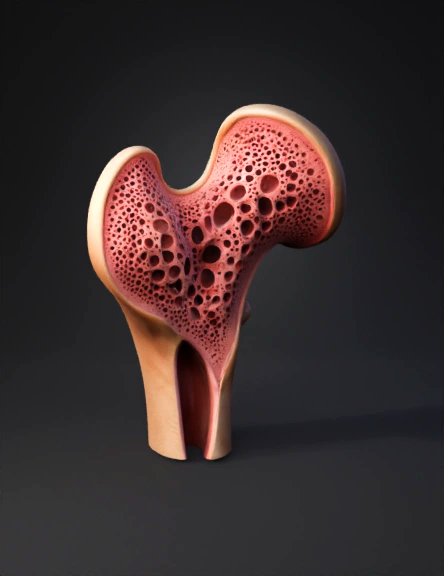
대퇴골 해면골 3d 모델 3D Model
Femur Bone 3D Model
Fémur Modelo 3d 3D Model