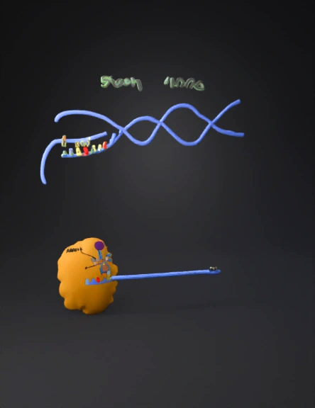
Biology Diagram 3D Model
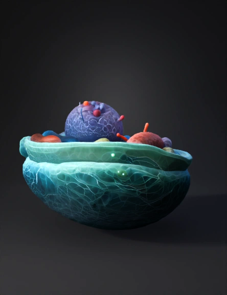
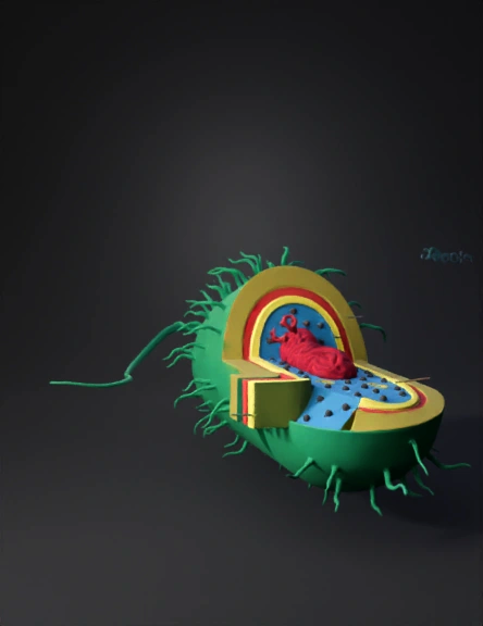
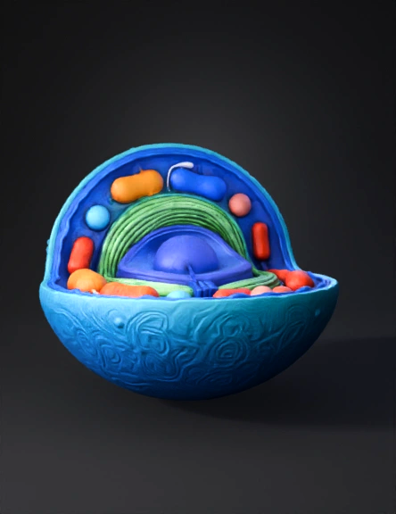
Eukaryotic Cell 3D Model
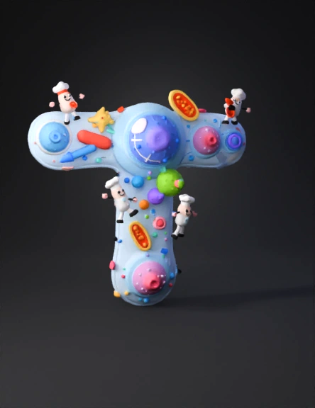
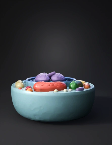
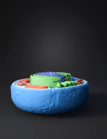
Whimsical Cell Model 3d 3D Model
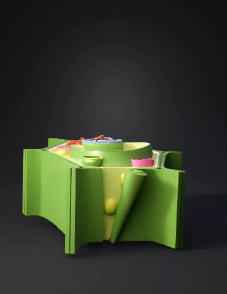
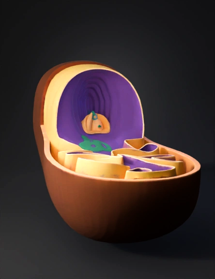
Modelo 3d De Célula Vegetal 3D Model
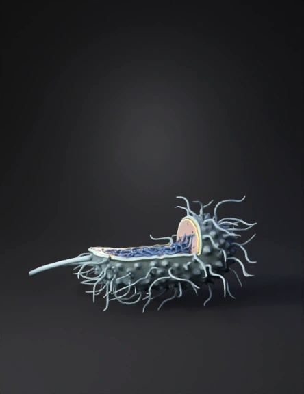
Modelo 3d De Bacteria 3D Model
Bacterial Cell 3D Model
Célula Animal 3d 3D Model
Bacterial Cell 3D Model
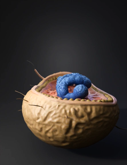
Mitochondrion 3D Model
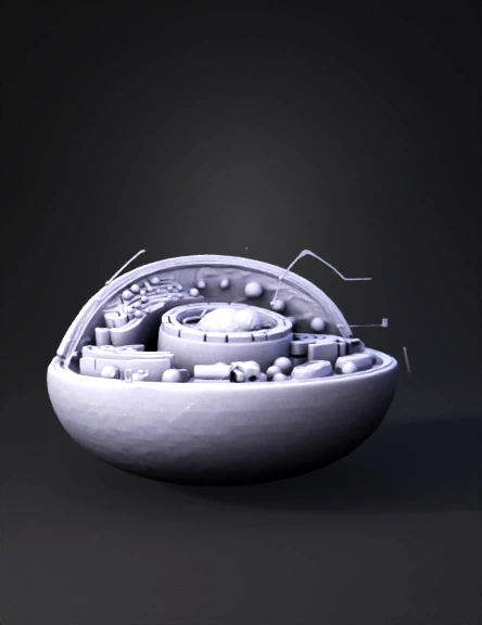
Cell Diagram 3D Model
Mitochondrion 3D Model
Cell Anatomy Diagram 3D Model
Animal Cell 3D Model
Mitochondrion 3D Model
Cell Diagram 3D Model
Modelo 3d De Célula Animal 3D Model
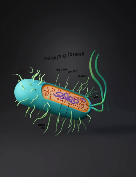
Bacterial Cell 3D Model
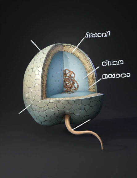
Thermococcus Archaeon 3D Model