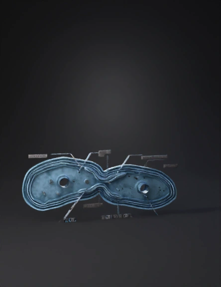
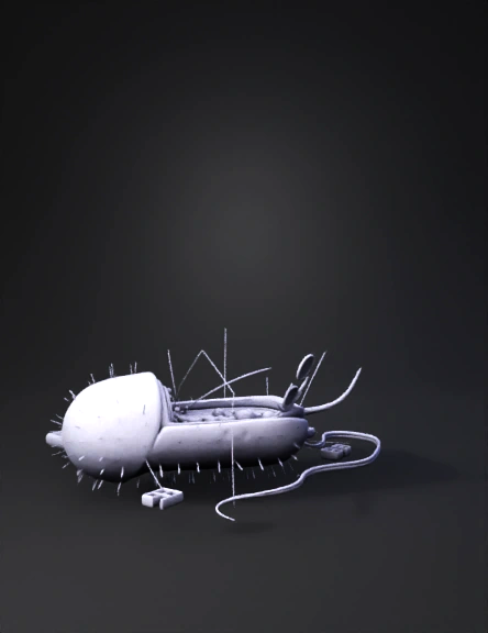
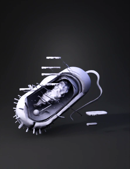
Bacterial Cell 3D Model
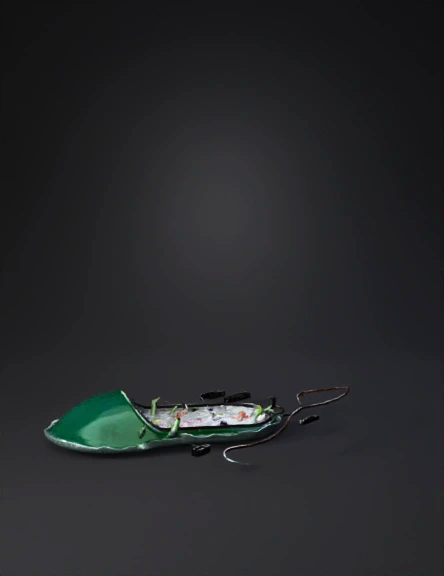
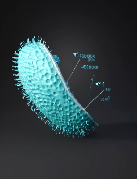
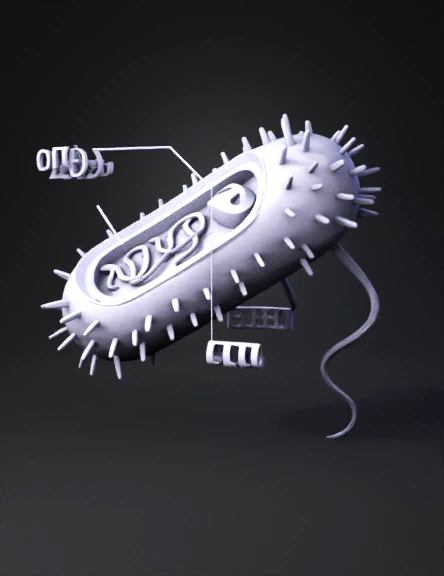
Bacterium 3D Model
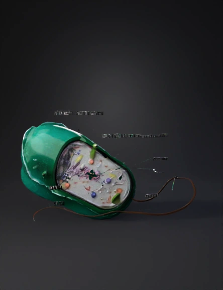
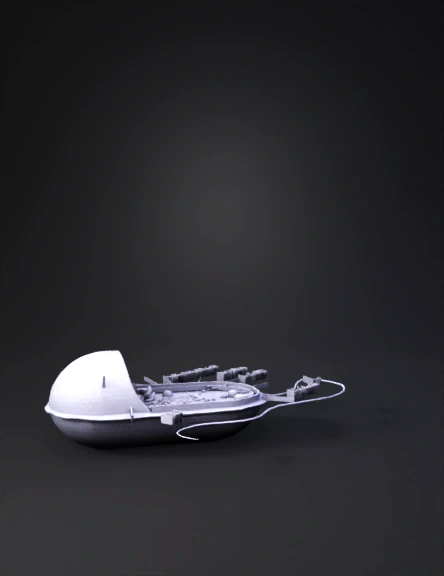
Bacteria 3D Model
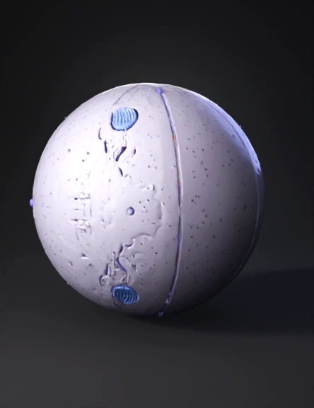
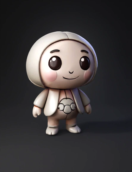
Animal Cell 3D Model
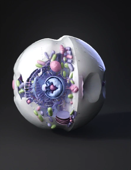
Animal Cell 3D Model
Bacterial Cell 3D Model
E3cbaee1-aa6f-40cd-be55-37f925651bb7 3D Model
0c1a3a44-8c36-4700-8094-0b7b064f8246 3D Model
120a543f-2525-45c3-a946-53ad8f886bb6 3D Model
Edddb4a6-6f25-4a5b-a9f4-89031911ee43 3D Model
B999a920-0f07-4c06-9422-dc86b4fbf332 3D Model
Dbddf595-27bc-484c-b6c7-ea71d909b160 3D Model
A2ee2de3-982f-49ba-a23e-52403006ea1e 3D Model
6d9cdd27-aea6-4d0b-b667-d7e558a5d2ef 3D Model
30d296e9-9e29-468c-a821-b1a5c253f3d3 3D Model
B0a41e0e-b1f7-4a3c-819c-ff64592083b3 3D Model
465adb99-95bb-4034-bf94-e6181dad68f3 3D Model
2dbd2236-69ef-42b6-b89b-9f0600548356 3D Model