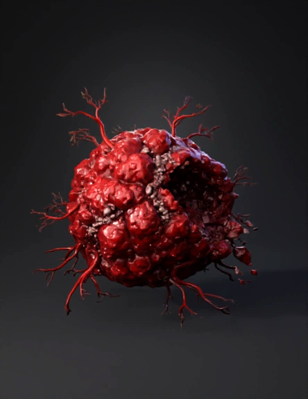
Tumor Cell Cluster 3D Model
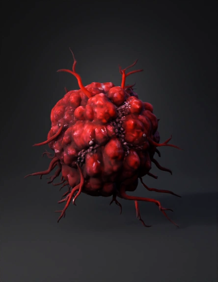
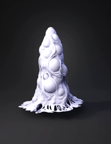
Organic Tumor 3D Model
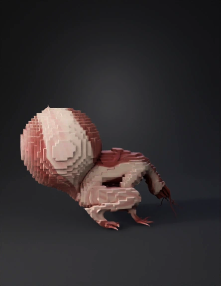
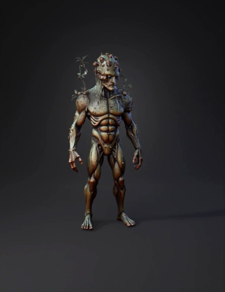
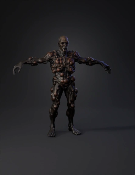
Tumor Creature 3D Model
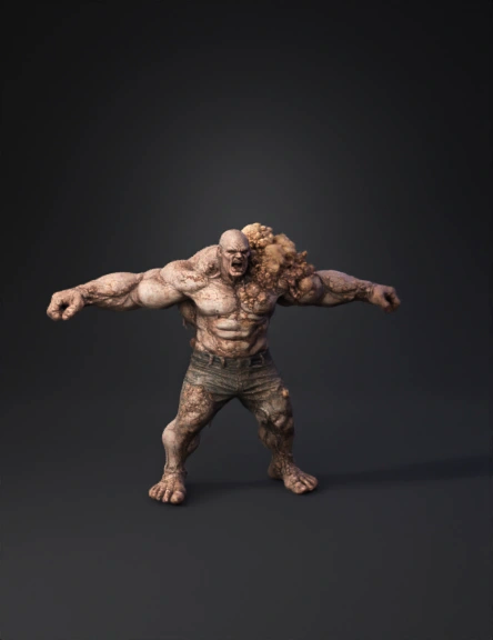
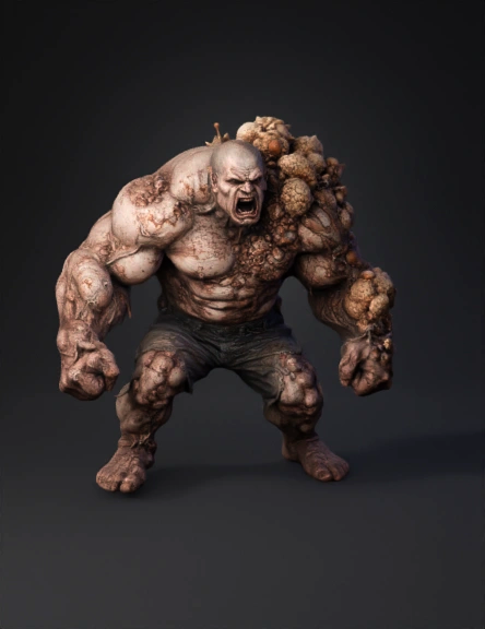
Grotesque Monster 3D Model
Horror Creature 3D Model
Grotesque Creature Head 3D Model
Organic Tumor 3D Model

Bone With Tumor 3D Model
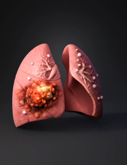
Lung Pair 3D Model
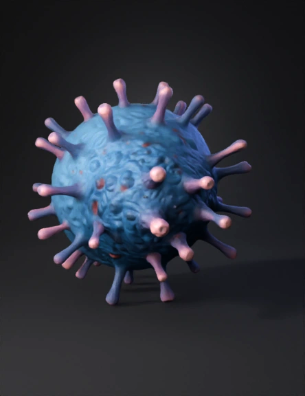
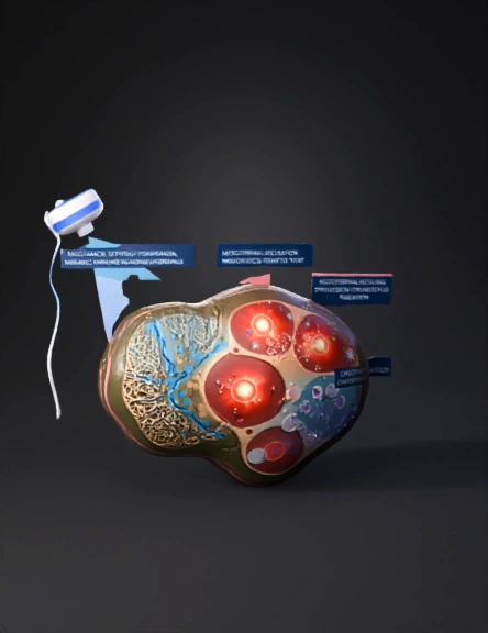
Cancer Cell 3D Model
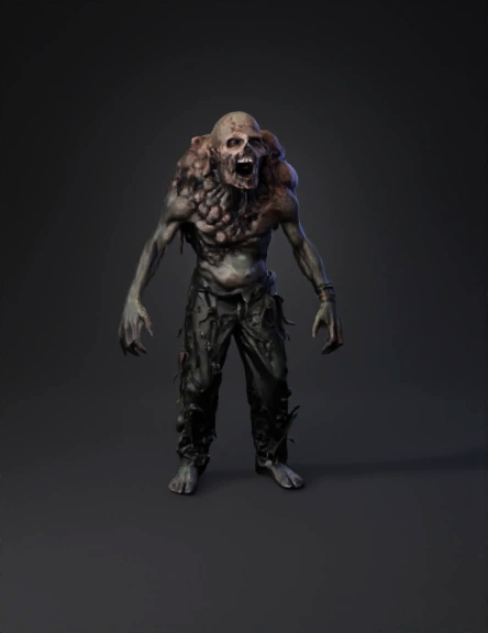
Monster 3D Model
Lung Tumor 3D Model
Abstract 3D Model
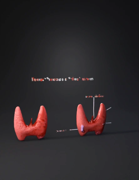
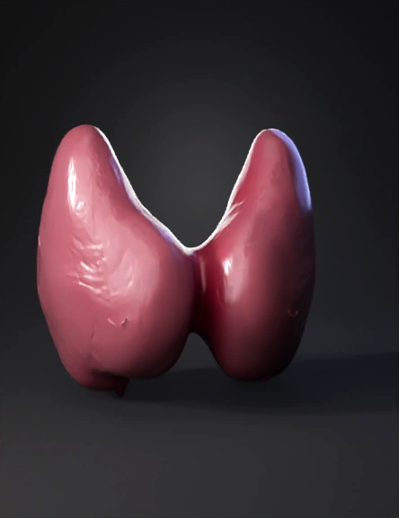
Thyroid Gland 3D Model
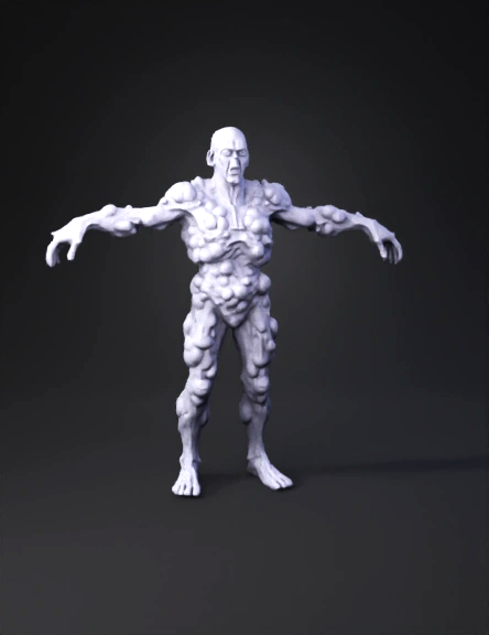
Monstrous Humanoid 3D Model
Unknown 3D Model
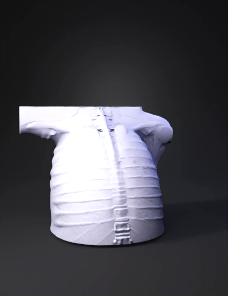
Skin Cross-section 3D Model
Oral Tumor 3D Model