3D Workspace
Home
Assets
Affiliate Program
Sign up/Log in
?
Upgrade
DCC Bridge
Anonymous1752063623
12-27 17:54
Model Name
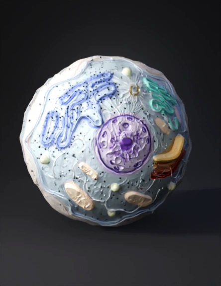
cell model 3d
Tags
biology
cell
nature environment
nature environment realistic
nature environment rendering
nature environment rendering realistic
organelles
realistic
rendering
rendering realistic
Prompt
🎯 GOAL Generate a scientifically accurate 3D model of an animal cell for secondary school biology education, with all major organelles, rendered in distinct realistic colors, and proportions based on peer-reviewed cell biology references. 🧬 SCIENTIFIC ACCURACY (CRITICAL – NO CARTOON STYLE) Cell type: Typical eukaryotic animal cell (mammalian somatic cell) Overall cell diameter: 20–30 µm (average ≈ 25 µm) All organelles must follow biologically accepted size ranges from cell biology journals and textbooks (e.g. Alberts, Lodish, Junqueira). Correct spatial organization (organelles positioned as in real cytoplasm, not symmetrically arranged). No plant organelles (no cell wall, no chloroplast, no large central vacuole). 🔬 ORGANELLES TO INCLUDE (MANDATORY) 1. Plasma Membrane Thickness: 7–10 nm Semi-transparent phospholipid bilayer Slight fluid curvature (not rigid) Color: translucent light beige / pale blue 2. Cytoplasm Gel-like texture Slight granularity Neutral light blue-grey tone 3. Nucleus Diameter: 5–10 µm (≈ 1/3 of cell diameter) Double nuclear envelope with visible nuclear pores Inner contents: Chromatin (diffuse, not chromosomes) Nucleolus (1–2 µm) Colors: Nuclear envelope: pale purple Chromatin: darker violet Nucleolus: deep purple 4. Rough Endoplasmic Reticulum (RER) Flattened membrane sacs Continuous with nuclear envelope Ribosomes attached (small dots) Color: blue-violet with dark ribosomes 5. Smooth Endoplasmic Reticulum (SER) Tubular membrane network No ribosomes Color: light cyan 6. Ribosomes Size: 20–30 nm Free ribosomes scattered in cytoplasm Bound ribosomes on RER Color: dark blue or black dots 7. Golgi Apparatus 4–8 flattened cisternae Slight curvature Positioned near nucleus Color gradient: orange → red 8. Mitochondria Length: 1–10 µm Double membrane Clearly visible cristae Several mitochondria distributed in cytoplasm Color: red / orange with darker inner folds 9. Lysosomes Diameter: 0.1–1 µm Spherical Acidic interior texture Color: yellow-green 10. Peroxisomes Diameter: 0.1–1 µm Smaller than lysosomes Color: light green 11. Centrosome Two perpendicular centrioles Microtubule organization visible Color: dark yellow / gold 12. Cytoskeleton (simplified but correct) Microtubules Actin filaments Intermediate filaments Color: faint white / light grey lines 📐 PROPORTIONS & SCALE (VERY IMPORTANT) Maintain relative size accuracy between: nucleus vs cell mitochondria vs lysosomes ribosomes vs membranes Use micrometer-based biological scaling, not artistic scaling. Include scale bar (µm) or internal reference. 🎨 VISUAL STYLE Hyper-realistic scientific 3D render Clean educational look No exaggerated colors High contrast between organelles Soft neutral lighting White or transparent background No labels in final render (labels added later externally) 🧪 EDUCATIONAL CONTEXT Audience: Secondary school students (ages 13–18) Purpose: Interactive learning, 3D rotation, zooming Must be suitable for: Biology textbooks Interactive simulations AR / VR educational use 🚫 STRICTLY AVOID Cartoon style Symmetrical organelle layout Incorrect organelle sizes Oversized mitochondria or ribosomes Plant-cell features Decorative or artistic distortion 📦 OUTPUT REQUIREMENTS Fully closed 3D mesh Clean topology Separate organelles as individual objects Ready for: Blender Unity / Unreal Web-based 3D viewers (GLTF / FBX / OBJ)
Detailed Info
Related Models
Enter invite code
Enter invite code to get credits!