3D Workspace
Home
Assets
Affiliate Program
Sign up/Log in
?
Upgrade
DCC Bridge
Anonymous1762543416
11-15 00:12
Model Name
horse anatomy 3d model
Tags
cranium
creatures animals
creatures animals realistic
creatures animals rendering
creatures animals rendering realistic
horse anatomy
realistic
rendering
rendering realistic
Prompt
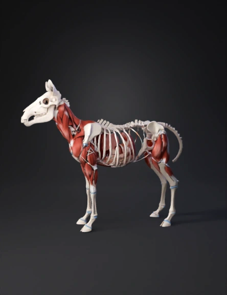
1.1. Skull (30–35 cm length) Elongated cranium with clearly defined nasal cavity, eye sockets, and jaw joints. Upper jaw (maxilla) and lower jaw (mandible) modeled as clean separate bone units. Nasal bones form a long, smooth arch forward. Cheekbones (zygomatic arches) form lateral support. 1.2. Vertebral Column 7 cervical vertebrae – long, curved, allowing neck mobility. 18 thoracic vertebrae – tall dorsal spines that support shoulder height. 6 lumbar vertebrae – wide, strong, supporting the abdomen. 5 fused sacral vertebrae – forming the pelvis attachment. 18–22 caudal vertebrae – tapering in size along the tail. 1.3. Ribcage 18 pairs of ribs, smooth curvature, forming an oval thoracic cavity. 1.4. Forelimbs Large scapula, angled backward. Humerus short and thick. Radius and ulna nearly fused. Metacarpal and phalangeal bones forming a single hoof support. 1.5. Hindlimbs Strong femur, long tibia, reduced fibula. Metatarsal bones elongated. Single hoof phalanx. 2. ARTICULAR TISSUES – SEPARATED FROM BOTH BONES AND MUSCLES 2.1. Cartilage Smooth, semi-transparent caps on all joint surfaces (clean, even thickness). Present on shoulders, elbows, knees, hocks, fetlocks, and jaw joints. 2.2. Ligaments White, ribbon-like structures stabilizing each joint. Distinct groups: Shoulder suspensory ligaments Elbow collateral ligaments Knee and hock ligaments Intervertebral ligaments connecting vertebrae 2.3. Tendons Long, glossy tendon bands on limbs, especially forelimb flexor and extensor tendons. Clear separation between flexor tendon sheaths and extensor tendons. 3. MUSCULAR SYSTEM – ALL MUSCLE GROUPS ISOLATED AND SEPARATE 3.1. Neck Muscles Superficial: Brachiocephalicus: long ribbon muscle from head to shoulder. Sternocephalicus: runs from sternum to lower jaw. Deep: Splenius, longus colli, multifidus cervicis arranged in layered segments. 3.2. Trunk Muscles Superficial layer: Trapezius: triangular sheet over the scapula. Latissimus dorsi: large lateral muscle. Mid-layer: Longissimus dorsi: long, thick muscle mass flanking the spine. Serratus ventralis: fan-like muscle connecting ribs to scapula. Abdominal group: Rectus abdominis: paired long muscles along the belly. External & Internal obliques: angled sheets forming side walls. 3.3. Forelimb Muscles Deltoid: rounded mass on the shoulder. Triceps brachii: thick multi-headed muscle of upper forelimb. Extensor group: long, thin muscles for limb extension. Flexor group: deeper muscles controlling the hoof area. 3.4. Hindlimb Muscles Gluteal region: Gluteus medius: major, dome-shaped hindquarter muscle. Gluteus superficialis: covers upper hip contour. Hamstring group: Biceps femoris: large and lateral. Semitendinosus: cylindrical, posterior. Semimembranosus: broad and medial. Deep: Quadriceps femoris: powerful knee extensor. Gastrocnemius: primary calf muscle. 4. SUBCUTANEOUS TISSUE (WITHOUT SKIN) A thin, clean layer representing soft connective/fat tissue. Smooth, uniform, slightly elastic. Lies between muscles and where skin would be, but the skin layer is removed. 5. INTERNAL ORGANS – SEPARATE, CLEAN, AND CLEARLY SHAPED 5.1. Heart Cone-shaped organ positioned behind the forelimbs. Clean separation from surrounding structures. 5.2. Lungs Two large, soft lobes. Smooth outer surfaces, symmetric. 5.3. Liver Large, multi-lobed, smooth contour. 5.4. Digestive Organs Stomach: single-chambered, rounded. Small and large intestines: long tubular loops arranged in a bundle. 5.5. Kidneys Two bean-shaped organs placed symmetrically. 6. MOUTH, TEETH, TONGUE — DETAILED, CLEAN, NON-GRAPHIC, FULLY SEPARATED 6.1. Upper and Lower Jaws (Skeletal) Maxilla and mandible clearly visible without skin. Dental arches cleanly exposed. 6.2. Teeth Zebras are grazers, so their teeth are specialized: Incisors (Front teeth) Six upper and six lower. Wide, flat surfaces for cropping grass. Arranged in a neat horizontal arc. Canines Present but small (often reduced). Premolars and Molars Large, ridged chewing surfaces. Organized into two symmetrical upper and lower rows. Back teeth form a wide grinding platform. All teeth appear clean, ivory-colored, hard-surfaced, and neatly aligned. 6.3. Tongue Long, muscular, flexible organ. Smooth exterior surface, no skin, no papilla depiction. Slightly tapered tip. Anchored firmly to the hyoid bones inside the throat. Presented as a separate anatomical object for modeling. 6.4. Palate and Mouth Cavity Hard palate: ridged bony roof of the mouth. Soft palate: smooth muscular sheet at rear. Clear separation from jaw bones, teeth, tongue, and throat.
Detailed Info
Related Models
Enter invite code
Enter invite code to get credits!