3D Workspace
Home
Assets
Affiliate Program
Creator Program
Sign up/Log in
View Plans
DCC Bridge
natalia.carvalhoalves
11-16 18:09
Model Name
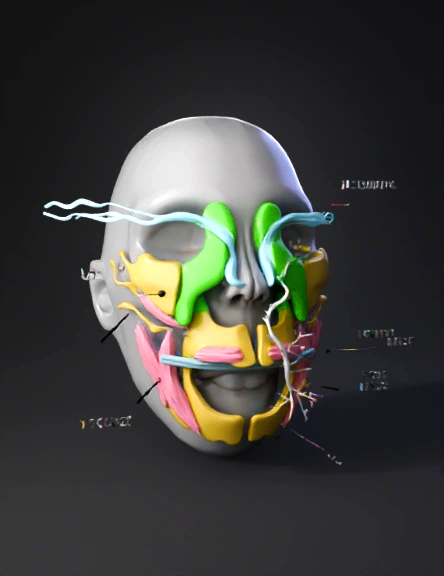
5-week embryo 3d model
Tags
3d printing
3d printing realistic
embryo
props
props 3d printing
props 3d printing realistic
props realistic
realistic
Prompt
A highly detailed 3D embryology model of human facial development at 5 weeks gestation, focusing on the first pharyngeal arch and neural crest cell migration. Base: Oval embryonic head (light gray) with 5 visible pharyngeal arches marked by shallow grooves on both sides. Highlight the 1st pharyngeal arch (bright green) bilaterally: - Divided into maxillary process (upper, thinner, pointing toward midline) - Mandibular process (lower, wider, forming U-shape) Neural crest cells: Represented as glowing blue filaments/streams migrating from the dorsal neural tube into the green 1st arch. Derived structures from 1st arch (color-coded, semi-transparent to show origin): - Bones (yellow): mandible (U-shaped), maxilla (paired plates), zygomatic bones, temporal bone (squamous part), malleus and incus (tiny ossicles near ear region) - Muscles (pink): masseter, temporalis, pterygoids (as fiber bundles on mandible) - Nerve (white): trigeminal nerve (CN V) branching into V1, V2, V3 from Meckel’s cartilage - Cartilage (light blue): Meckel’s cartilage rod inside mandibular process Include small floating labels (white text on black pins) for: "1st Pharyngeal Arch", "Neural Crest Cells", "Maxillary Process", "Mandibular Process", "Mandible", "Trigeminal Nerve". Add a subtle red arrow showing migration path of neural crest cells into the arch. Style: Scientific illustration, clean, colorful, medical accuracy, smooth surfaces, high detail, studio lighting, white background. Optimized for 3D printing and educational use.
Detailed Info
Related Models
Enter invite code
Enter invite code to get credits!
 natalia.carvalhoalves


 natalia.carvalhoalves