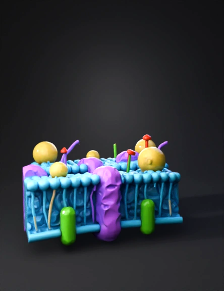
cell membrane 3d model
A simplified model of the cell membrane (Cell Membrane - Fluid Mosaic Model), with its anatomical structure detailed as follows: 1. Phospholipid Bilayer: Color: Cyan/Light Blue. Description: Two parallel layers; each layer consists of tightly packed rows of small spheres (representing hydrophilic heads) with filaments extending inwards (representing hydrophobic tails). The two layers are opposite each other at the tail end. 2. Integral/Transmembrane Proteins: Color: Magenta/Purple. Description: Large structures with a wavy texture that penetrate the two layers of the blue membrane lengthwise from top to bottom. These represent protein channels or receptors in the cell. 3. Additional components and receptors: Yellow sphere: A bright yellow, spherical structure located on the outer surface of the membrane (representing a peripheral protein or a lipid molecule). Green cylinder: A neon/light green, cylindrical structure that penetrates the lower part of the membrane horizontally/obliquely (possibly representing c
4K·PBR·v3.1-20260211


