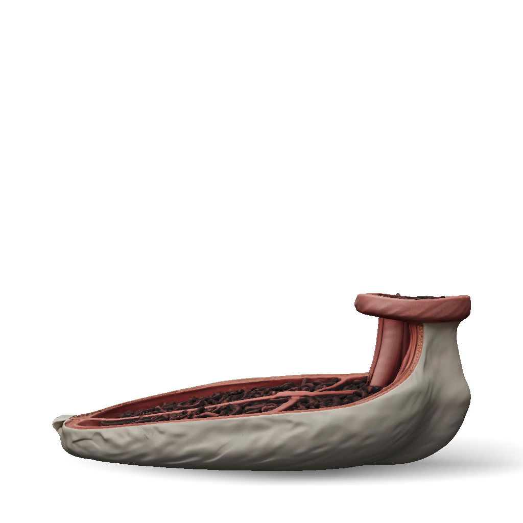
bovine rumen 3d model
Anatomical 3D model of a bovine rumen shown in cutaway cross-section, half of the organ removed to reveal the internal wall structure. The internal surface of the rumen wall is densely covered with numerous finger-like, leaf-shaped papillae (ruminal papillae), varying in density: very dense and long papillae in the ventral sac, moderately dense in the caudal blind sacs, sparse and short papillae in the dorsal sac, and completely smooth, papillae-free surface on top of the muscular pillars (dorsal pillar, ventral pillar, right and left longitudinal pillars, cranial and caudal pillars), which appear as thick raised smooth bands crossing the internal wall. Internal mucosa color: dark brown to blackish-brown, velvety texture from the dense papillae, in contrast to the pale pink-gray external serosal surface. Cut edge shows wall thickness with distinguishable layers: mucosa (papillated), submucosa, two muscular layers, and outer serosa. Style: realistic veterinary anatomical te
4K·PBR·v3.1-20260211


