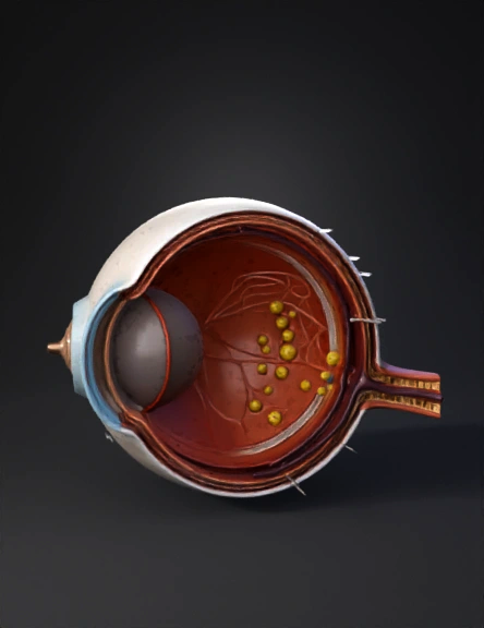
eye anatomy 3d model
Create a highly detailed 3D medical visualization of a human eye affected by diabetic retinopathy. The eye should be shown in cross-section to clearly display internal anatomical structures, including the cornea, lens, vitreous humor, retina, optic nerve, and blood vessels. Focus on the retina showing visible pathological changes associated with diabetic retinopathy: microaneurysms, dot and blot hemorrhages, hard exudates (yellow lipid deposits), cotton wool spots, retinal swelling, and areas of neovascularization with fragile, abnormal blood vessel growth. The style should be hyper-realistic, medically accurate, and suitable for an ophthalmology textbook or clinical presentation. Use dramatic but clinical lighting to highlight damaged retinal areas. The texture should show subtle inflammation and vascular leakage. Render in high resolution (4K), photorealistic materials, subsurface scattering for organic tissue, and sharp anatomical detail. Neutral background, studio lighting, scientific visualization style
4K·PBR·v3.0-20250812


