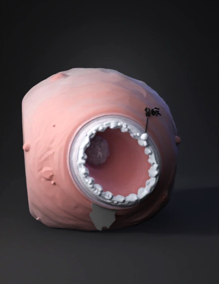
blastocyst 3d model
Create a photorealistic, scientifically accurate 3D animated sequence depicting human embryo implantation (Days 6–12 post-fertilization), based on established embryological references (Moore & Persaud, Gray's Anatomy). ═══════════════════════════════════════════════════════════ SCENE 1 — BLASTOCYST ARRIVAL (Day 6) ═══════════════════════════════════════════════════════════ Model a free-floating blastocyst (~0.1–0.2 mm diameter) approaching the posterior wall of the uterine fundus. Blastocyst anatomy must include: - Zona pellucida: translucent, glycoprotein shell — shown in active dissolution (hatching) via enzymatic lysis - Trophoblast layer: single outer epithelial ring of cuboidal cells (cytotrophoblast), slightly irregular surface texture, pale eosinophilic tone - Inner Cell Mass (embryoblast): compact, eccentric cluster of darker, tightly-packed cells positioned toward the endometrial surface - Blastocoel: fluid-filled central cavity, optically clear with slight
4K·v3.1-20260211


