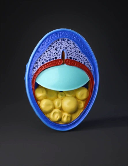
frog embryo 3d model
Create a scientifically accurate 3D model of an early gastrula stage frog embryo (Xenopus laevis). The embryo should be shown as a hemispherical cross-section, cut vertically through the animal-vegetal axis to reveal the internal structure. The model should clearly display: Animal pole at the top, composed of small pigmented cells Vegetal pole at the bottom, composed of large yolk-rich cells Blastocoel cavity in the upper hemisphere Dorsal lip of the blastopore forming on one side Involuting mesoderm cells migrating inward beneath the ectoderm Archenteron beginning to form as a small cavity Distinct germ layers: ectoderm (outer layer), mesoderm (middle migrating layer), and endoderm (large yolky inner cells) Use educational colors: Ectoderm: blue Mesoderm: red Endoderm: yellow Blastocoel and archenteron: transparent light cyan The cut surface should be smooth and clearly expose internal anatomy. The external surface should remain rounded and realistic. Style should be clean, high-
4K·PBR·v3.1-20260211


