3D Workspace
Home
Assets
Affiliate Program
Creator Program
Sign up/Log in
View Plans
DCC Bridge
xarehi6978
11-05 06:16
Model Name
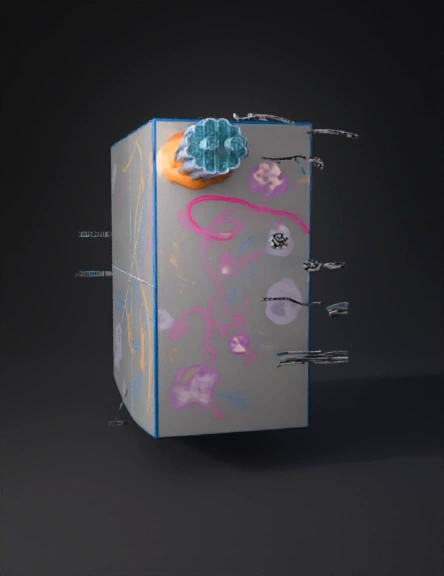
connective tissue 3d model
Tags
connective tissue
nature environment
nature environment realistic
nature environment simulation
nature environment simulation realistic
realistic
simulation
simulation realistic
Input
Prompt
Generate a highly detailed, anatomically accurate 3D model of connective tissue from this 2D diagram. The model must include: 1. A transparent 3D cube representing the extracellular matrix, with visible depth and spatial arrangement. 2. All labeled cell types in their correct positions and proportions: - Fibroblasts (elongated, spindle-shaped, with nuclei) - Macrophages (amoeboid shape, irregular membrane) - Lymphocytes (small, round, dense nucleus) - Plasma cells (eccentric nucleus, abundant cytoplasm) - Neutrophils (multi-lobed nucleus, granular cytoplasm) 3. Structural fibers accurately rendered: - Collagen bundles (thick, wavy, rope-like, pink/orange) - Elastic fibers (thin, branching, yellow) - Reticular fibers (fine, mesh-like, purple) 4. Vascular and neural elements: - Capillary (red, tubular, endothelial lining visible) - Nerve fiber (yellow, cylindrical, with myelin sheath if possible) 5. Include subtle surface textures on cells (e.g., microvilli or rough ER in plasma cells). 6. Maintain scientific accuracy: cells should not overlap unnaturally; fibers should interweave realistically. 7. Output format: .glb or .obj, ready for download and viewing in 3D software (Blender, Windows 3D Viewer, etc.). 8. Style: Medical illustration realism — clean, educational, no artistic exaggeration. Note: This is for educational use in histology. Prioritize clarity and biological fidelity over artistic flair.
Detailed Info
Related Models
Enter invite code
Enter invite code to get credits!
 xarehi6978


 xarehi6978