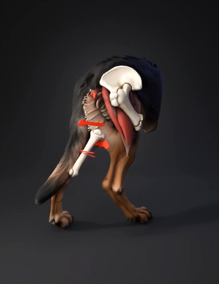
canine hind leg 3d model
Generate a highly detailed, anatomically precise 3D rendering of the right hind leg skeletal system of an adult German Shepherd dog, isolated on a clean white background, highlighting a mid-diaphyseal oblique fracture of the femur. Depict the fracture as a clean, displaced break in the central shaft of the femur (thigh bone), with the distal segment shifted caudoproximal due to gastrocnemius and hamstring muscle pull, showing jagged bone ends and subtle hematoma swelling. Include surrounding bones clearly: pelvis proximally, straight foreleg-like parallel femur with head, neck, greater trochanter; tibia, fibula, tarsus distally; realistic bone textures with cortical thickness, trabecular interiors visible at the break, soft shadows for depth, and red arrows labeling 'Femur Fracture Site' and muscle pull directions. Use a side profile (lateral view) pose in a scientific veterinary illustration style for educational use, high resolution.
4K·v3.1-20260211


