3D Workspace
Home
Assets
Affiliate Program
Sign up/Log in
?
Upgrade
DCC Bridge
Anonymous1766825207
12-27 09:34
Model Name
human body 3d model
Tags
character
character realistic
character rendering
character rendering realistic
human body
medical
person lying down
realistic
rendering
rendering realistic
Input
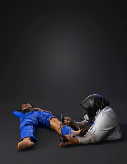
Prompt
Ultra-high detail 3D medical illustration depicting a complete and correct Ankle Brachial Index (ABI) examination, with strong emphasis on the examiner actively using both a sphygmomanometer and Doppler ultrasound device. A female healthcare professional is clearly visible and visually dominant in the scene. She wears a clean white medical coat, long-sleeve clinical attire, and a black hijab, representing a trained medical examiner. Her upper body, arms, hands, and face are fully visible, with a focused and professional facial expression, eyes directed toward the Doppler device and pressure gauge. The examiner is positioned kneeling beside the patient, demonstrating correct ergonomics during ABI measurement. Right hand: firmly holding a handheld Doppler ultrasound probe, with ultrasound gel visibly applied to the probe tip and the patient’s skin. The probe is accurately positioned at a 45–60 degree angle over the posterior tibial artery, precisely located posterior to the medial malleolus. Left hand: actively stabilizing the patient’s foot and ankle while simultaneously controlling the inflation bulb of the sphygmomanometer, clearly showing the examiner is inflating and deflating the cuff while listening to Doppler sounds. A properly sized blood pressure cuff is wrapped snugly around the distal lower leg just above the ankle, with visible tubing connected to an analog sphygmomanometer gauge, which is readable and facing the examiner. The male patient is lying in a supine position on a clean, light-gray clinical tiled floor, relaxed and cooperative, one leg extended and slightly externally rotated to expose the ankle anatomy. Anatomical precision is emphasized using semi-transparent, educational overlays that clearly illustrate: Posterior Tibial Artery (highlighted behind the medial malleolus) Dorsalis Pedis Artery (highlighted on the dorsum of the foot between the first and second metatarsals as an alternative ABI measurement site) Thin instructional arrows, labels, and subtle glowing outlines guide the viewer’s attention to the exact probe placement, cuff position, and arterial landmarks. The scene clearly communicates the clinical sequence of ABI measurement: cuff inflation, Doppler signal detection, and pressure reading. Lighting is bright, neutral, and clinical, eliminating shadows and ensuring maximum visibility of the examiner’s hands, medical devices, tubing, pressure gauge, and Doppler probe orientation. Style: hyper-realistic, professional 3D medical education illustration, photorealistic skin textures, accurate anatomy, realistic medical equipment proportions, no cartoon or stylized appearance. Camera angle: slightly elevated three-quarter side view, intentionally framed to keep the examiner’s body, hands, Doppler device, cuff, and ankle region all within the primary focal plane, making the examiner’s role unmistakably clear. Render quality: 8K resolution, ultra-sharp focus, realistic fabric folds, clear device markings, suitable for OSCE stations, clinical skills textbooks, anatomy teaching posters, and medical presentations.
Detailed Info
Related Models
Enter invite code
Enter invite code to get credits!