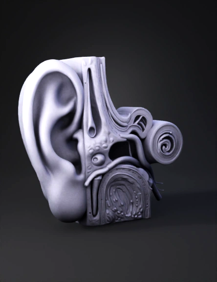
ear anatomy 3d model
Ultra‑realistic 3D anatomical model of the human ear, single‑sided continuous structure with no duplication or mirrored halves. Outer ear includes auricle (pinna) and external auditory canal leading to tympanic membrane. Middle ear cavity contains ossicles (malleus, incus, stapes) surrounded by yellowish bone tissue, with blood vessels in red and veins in blue. Inner ear features spiral cochlea, semicircular canals, vestibule, and auditory nerve fibers extending into temporal bone. Cross‑sectional view reveals surrounding bone, cartilage, and connective tissue with lifelike textures. Ensure nerves and vessels are clearly depicted entering and exiting ear region. Lighting emphasizes contrast between bone, soft tissue, and vascular structures. Composition must replicate exact proportions, coloration, and fine anatomical detail for medical visualization, strictly one‑sided without mirrored duplication.
v3.1-20260211


