3D Workspace
Home
Assets
Affiliate Program
Sign up/Log in
?
Upgrade
DCC Bridge
Anonymous1764233479
11-27 08:53
Model Name
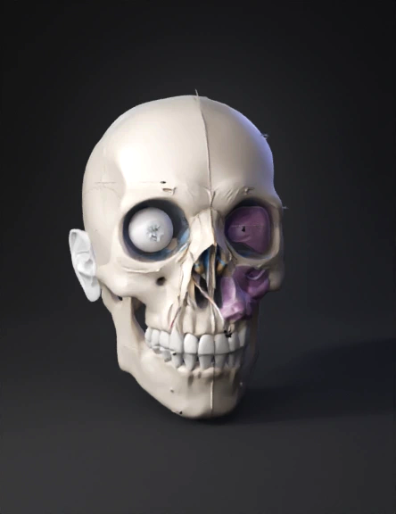
head 3d model
Tags
anatomical head
head
props
props realistic
props rendering
props rendering realistic
realistic
rendering
rendering realistic
Prompt
You are a senior medical imaging & 3D modeling engineer working for the FaceOpS / ALINA platform. Your task: generate a high-fidelity 3D model from a CT Head (non-contrast) DICOM study that matches, as precisely as possible, the following ultra-detailed radiological report and measurements. The model will be used in the “Imaging → 3D View” section of FaceOpS. SOURCE STUDY CONTEXT Location: Toshkent shahar klinikasi (Tashkent City Clinic) Study: CT Head series (non-contrast) Exam date: 05.11.2025 Patient: male, approx. 21 years old (DoB 01.01.2004) Acquisition: multidetector CT of paranasal sinuses + orbits + facial skeleton Slice thickness: 0.625 mm isotropic, bone + soft-tissue algorithm, 3D-VRT, multiplanar reformation. The user will upload the original DICOM series. You must: Ingest the CT DICOM. Perform segmentation of key anatomical and pathological structures. Generate clean, watertight 3D meshes (STL/OBJ/glTF) suitable for surgical planning and visualization. Attach metadata and measurements consistent with the report below. KEY DIAGNOSES TO REPRESENT IN 3D (FROM REPORT) Healed right medial orbital wall blow-out fracture (pure type) with residual deformity Codes: ICD-11 S02.31 + NB2.1 (AO CMF context) Description: Fracture line along right lamina papyracea (anterior + middle thirds), segment length 9–11 mm. Minimal medial displacement = 2.1 mm inward. Rounded margins → healed > 6 months. No entrapment of medial rectus or orbital fat herniation into ethmoid. 3D REQUIREMENT: Segment: right medial orbital wall, fracture line, surrounding bone. Represent the slight inward bowing (2.1 mm) of the lamina papyracea. Keep orbital contents (globe, muscles, fat) anatomically realistic with no herniation through the defect. Healed nasal bone fracture with secondary naso-orbital deformity Codes: ICD-11 S02.2 + Le Fort–Cummings class II Description: Comminuted nasal bones with leftward displacement of the entire nasal pyramid (5.7 mm at bony dorsum level). Overriding fragments, callus formation. Resultant saddle-nose + crooked nose deformity. 3D REQUIREMENT: Segment nasal bones and nasal pyramid. Show callus and residual deformity. Apply the quantified deviation: 5.7 mm left at rhinion/bony dorsum. High post-traumatic nasal septal deviation (S-shaped) Codes: ICD-11 DA0B.0, Killian Type IV Description: Septal deviation angle: 16° to the left at bony-cartilaginous junction + 11° to the right at caudal end → S-shaped deformity. Spurs at vomer–maxillary crest contact zone, blocking right middle meatus. Severe, combined bony + cartilaginous deviation. 3D REQUIREMENT: Segment septal cartilage + bony septum (vomer, perpendicular plate). Recreate the S-shaped deviation with angles ~16° and 11°. Visualize the spur near the vomer–maxillary crest and its relationship to the right middle meatus. Chronic rhinosinusitis with polyps – right-sided dominant Codes: ICD-11 CA0C.1 + EPOS 2020 stage 3 (CRSwNP) Description: Right maxillary sinus is completely opacified by a polypoid mass 22 × 18 × 26 mm arising from inferomedial wall (infundibular origin). LM score: 10/12 right, 4/12 left. Ostiomeatal complex completely obstructed by turbinate + deviated septum. Chronic mucosal thickening. 3D REQUIREMENT: Segment right maxillary sinus cavity and the soft-tissue polypoid mass as a separate structure. Approximate mass size and volume according to report: ~5.5 cm³ with dimensions 22 × 18 × 26 mm. Show complete obstruction of the sinus ostium and ostiomeatal complex on the right. Right inferior turbinate hypertrophy Codes: ICD-11 DA08.1 Description: Bony + mucosal hypertrophy of the right inferior turbinate. Occupies 78% of the right common nasal meatus on coronal plane. 3D REQUIREMENT: Segment inferior turbinates bilaterally. Make right inferior turbinate visibly enlarged so that, in coronal view, it narrows the airway and occupies ~78% of the right common meatus. No intracranial, globe, optic nerve, superior orbital fissure, or extraocular muscle pathology Normal Hertel index, no enophthalmos measurable on CT, optic canal intact. 3D REQUIREMENT: Basic representation of intracranial vault outline, globes, optic nerves, and extraocular muscles as “normal” reference structures, without pathologic deformation.
Detailed Info
Related Models
Enter invite code
Enter invite code to get credits!